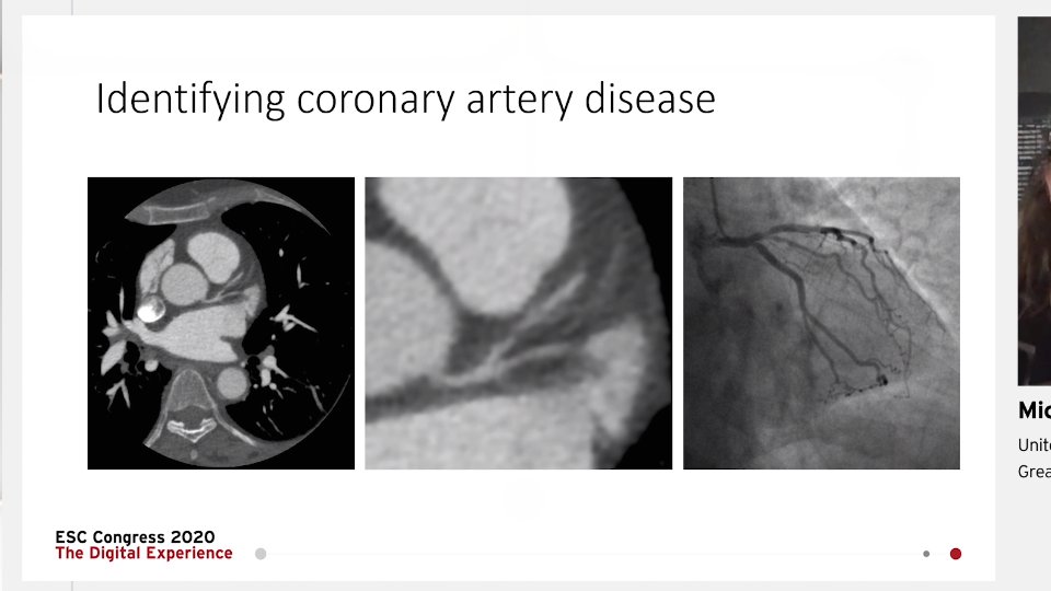
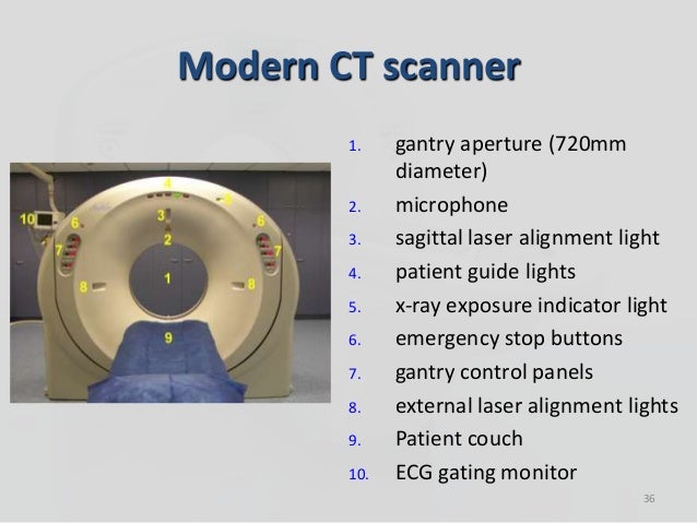
The micro-computed tomography (microCT) core provides high-resolution assessments of density, geometry and microarchitecture of mineralized tissues, such as bones and teeth, calcification as result of pathology, or soft tissues and biomaterials stained with radiographic contrast media. X-Ray Micro-Computed Tomography (μCT) X-ray micro-computed tomography (μCT) is a nondestructive imaging technique that allows for food microstructure to be characterized in the native state (Schoeman et al., 2016; Wang et al., 2018). During measurement, X-rays pass through the sample and are either absorbed or scattered.
Are you considering getting certified by taking the Computed Tomography (CT) exam? If so, know that your preparation is the most important piece to succeeding on test day! The CT test is a difficult exam that requires a lot of study and preparation in order to pass.
For this reason, we created our CT practice test to help test takers learn about what they will see on the actual test. Knowing the format and wording of the questions isn’t enough, you will also need to take a look at the layout and breakdown of the test to truly optimize your study habits, so let’s take a look! If you would like to check out some sample questions, check below!
The CT exam contains 185 multiple-choice questions of which twenty (20) are trial questions that do not count for or against you. The breakdown of the test is as follows:
- Patient Care and Safety (36 items)
- Imaging Procedures (75 items)
- Physics and Instrumentation (54 items)
Test takers will have 3 1/2 hours to complete the examination. All scores are scaled in a possible range of 1 to 99, and the minimum passing score is 75. With these things in mind, choose your study material and preparation time wisely to ensure you will be ready for the exam. If you do not know where to start, check out our free CT practice test that will give you an idea of the questions on the test.
Computed Tomography Exam Basics Review
Watch this video on YouTube
CT Study Guide
Mometrix Academy is a completely free resource provided by Mometrix Test Preparation. If you find benefit from our efforts here, check out our premium quality CT study guide to take your studying to the next level. Just click the CT study guide link below. Your purchase also helps us make even more great, free content for test-takers.
Upgrade your studying with our CT study guide and flashcards:
CT Study Guide
CT Flashcards
Computer Tomography Study Guide Customer Success Stories
Our customers love the tutorial videos from Mometrix Academy that we have incorporated into our Computer Tomography study guide. The Computer Tomography study guide reviews below are examples of customer experiences.The book seems to outline the IMPORTANT stuff. I LOVE that there are explanations with the answer key for the practice test. I feel it is very important to know why an answer is right if I choose wrong.
 Computer Tomography Study Guide – Devin
Easy to follow, useful information. I enjoy the way it's broken into smaller sections/headings so I'm not overwhelmed with a lot of information all at once, very organized.
 Computer Tomography Study Guide – Abby
I love it. Relevant information, well condensed and presented in a way that it can be easily understood.
Computer Tomography Study Guide – CaesarMic Computed Tomography Program
I like the fact that the book gives the short and straight to the point material. I would recommend this book to others studying for CT boards.
Computer Tomography Study Guide – SumnerI now have a better understanding of some of the material that we went over in class due to the review of the book. I highly recommend it.
Computer Tomography Study Guide – CustomerComputed Tomography Mic Book
I loved the study supplies. They were really great sources and had a lot of the information that was covered on the test and made me more relaxed when I took it.
Computer Tomography Study Guide – TamikoSince I have received the practice exam materials and the flash cards, my levels of assessments and awareness has improved tremendous. The advice and suggested ways of studying daily has afforded me the time and 'no excuse' in preparing for the state exam for the LCDC (Licensed Chemical Dependency Counselor). I had been searching for over two months for study materials that would help me and I stumbled across your Web Site. I am grateful and would highly recommend your materials because of the simplicity and thorough researched information of the course study. Mometrix goes above and beyond in helping others to succeed.
Computer Tomography Study Guide – PeterI'm really happy with the guide, It's very informative but not too wordy and boring like the others I've looked at. It's written and broken down, in a way that is extremely easy to remember things. This is by far the best book I have looked at!
Computer Tomography Study Guide – KendallComputed Tomography Angiography
I have purchased several other brands of CT studying material and books and this study guide so far exceeds every one of them. It is precise and to the point without any rambling or confusion.
Computer Tomography Study Guide – HectorI am happy to have found this test preparation book. It has given me a nice refresher on the information I needed for the exam. It is very helpful that the materials are separated into categories, subcategories and detailed explanations. My test anxiety is lessening the more I study this book. This book provides the real support I need to study and pass my exam.
Computer Tomography Study Guide – EricaThe study guide is ideal. I was impressed with the fact that it dealt with studying topics from the nervous system, which is a very important part of the anatomy of the body that drug impairs and alters. I am really glad I chose this book.
Fabulous study guide not just for self-study, but for use as a teaching-tool during clinical supervisions with Addiction Counselor Interns.
Computer Tomography Study Guide – CustomerI felt that the information for other sections went in-depth in a way that facilitated my learning and I feel more prepared as a result!
Computer Tomography Study Guide – MeganI feel more confident now that I am studying from this book. I like the way it categorizes subjects and topics and is easy to read and create an outline. My favorite part is that it explains why the answer is the answer on the practice test. This definitely has helped tremendously.
Computer Tomography Study Guide – AerinI like that it breaks the material down into categories, and uses simple language to help those studying better understand the material without having to sort through loads of jargon. It also breaks down the material into categories for each of the 12 Core Functions which is crucial to studying, learning the information, and passing the test!
I really appreciated the 'How to Overcome Test Anxiety' section, because I tend to get nervous and that really helped me to feel more able to cope with my anxiety before and during the test day. Additionally, I felt that the information for other sections went in-depth in a way that facilitated my learning and I feel more prepared as a result!
Computer Tomography Study Guide – MeganI really like how everything is separated into sections and highlights what is important. As they say it is “fluff” free!
Computer Tomography Study Guide – JenniferI love this book; it is easy to read and carry around. I feel like I have learned so much more about substance abuse. I am so thankful I purchased this study material.
Computer Tomography Study Guide – Customer Mometrix Academy – Home
Introduction
The micro-computed tomography (microCT) core provides high-resolution assessments of density, geometry and microarchitecture of mineralized tissues, such as bones and teeth, calcification as result of pathology, or soft tissues and biomaterials stained with radiographic contrast media. Studies performed by the HSS microCT core include:
- density and architecture analysis of human biopsies (Fig.1)
Computed Tomography Quizlet
Figure 1: Human iliac crest biopsies
- phenotype characterization of bones and teeth in animal models (Fig. 2)
Adult mouse femur Mouse knee joint 7 month-mouse molar
Figure 2: Phenotype characterization of bone and teeth
- studies of particle-induced osteolysis (Fig. 3)
Figure 3: Mouse calvaria: particle-induced osteolysis
Computed Tomography Programs
- characterization of dystrophic calcifications
- visualization of bone implants
- biomaterial characterization
- vascular mapping
Equipment
Scanner: Scanco Medical MicroCT 35 (Fig. 4)
- Resolution: 1.75-38 microns. In a typical mouse bone characterization the resolution used is 6µm (for femur/tibia) or 10µm (for knee).
- Wide range of KVp (45-70)/selection of different filters, allowing for high- (soft/developing mineralized tissue), normal- (mature bone) and low-contrast (implants, tooth enamel) scans.
- Incremental scanning: Every field of view chosen for analysis is scanned by increments using a 2048 x 2048 CCD detector. This enables a precise choice of the analyzed area by the user.
- Batch scanning: Multiple specimens are loaded for scanning with pre-selected settings for efficient use of the system.
- Different setups for analysis of variable size samples
- Reproducibility of analysis: % range of measurements from repeated scans at a 6µm resolution:
- calibration phantom: <0.2% for mineral density
- mouse femur trabecular bone: <0.5% for mineral density, <0.7% for trabecular bone volume fraction, relative bone surface, trabecular thickness, number, and separation
Figure 4: Scanco Medical MicroCT 35
3D reconstruction and processing: HP Integrity server workstation.
The Scanco workstation combined with the 64bit-software for densitometry and histomorphometry analysis provides:
- archiving of scans
- fast 2D and 3D evaluation of reconstructed volumes
- automated density scaling correction
- automated beam hardening and center of rotation correction
- versatile definition of volume contour for analysis by user
- handling of very large datasets
- complete array of direct morphometry measurements in 3D
Core Support
The microCT core-associated staff meets with all new users before the beginning of analyses to discuss the technical options for the users’ purposes. The focus is on an efficient use of the system within the study budget. A limited amount of pilot analyses is also available for first-time studies.
Fee Schedule
- Scanning:
$75/hour (NIH-funded projects)
$100/hour (academic, non NIH funded)
$200/hr (corporate) - Analysis:
$75/hour (NIH-funded projects)
$100/hour (academic, non NIH funded)
$200/hr (corporate or other) when provided by the Core or $25/hour on the Core workstation by the user (training on analysis software is provided to users at the hourly rate).
Typical scanning time for mouse femurs: 6-10µm resolution ~1.5h (4 femurs can be scanned in parallel).
To schedule samples please review the Service Request Process and complete the form as provided below:
Service Request Process
Service Request Form
Mic Computed Tomography Registry Review
Contact Information
Kyung Hyun Park-Min, PhD
Assistant Scientist and Director
Email: ParkminK@HSS.edu
Hayat Benlarbi
Senior Research Technician
Email: BenlarbiH@HSS.
Phone: 212-774-7329
